 Just like a...

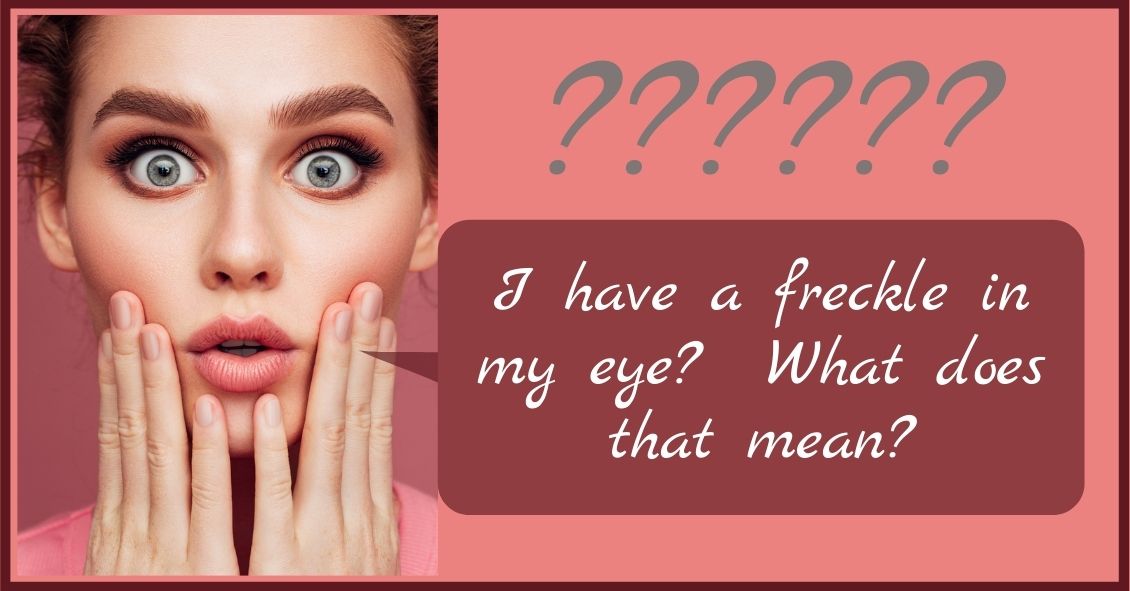
Choroidal nevus is the fancy term for a freckle in the back of the eye.
This lesion arises from a collection of cells that make pigment in the choroid, which lines the back of the retina and supplies the retina with nutrients. These choroidal nevi (plural of nevus) are usually grayish in color and develop in about 5-10% of the adult population. They are usually asymptomatic and detected during a routine dilated eye exam.
Just like any freckle on our body, we should monitor it for any change in size or growth. This is usually done with a photograph of the nevus and annual exams are normally recommended to monitor any change.
In addition to a photograph, other tests that can be used to monitor the nevus are:
- Optical coherence tomography - a test that uses light waves to take cross-section pictures of the retina. This test is used to detect if the nevus is elevated or if fluid is present underneath the retina.
- Ultrasound - uses sound waves to measure the size and elevation of the nevus.
- Fluorescein angiography - a dye test to detect abnormal blood flow through the nevus.
The concern is for transformation of the choroidal nevus into melanoma, a cancer in the eye. It has been estimated that 6% of the population have choroidal nevus and 1 in 8,000 of these nevi transform into melanoma. Some factors predictive of possible transformation in melanoma are:
- Thickness of the lesion, greater than 2 mm.
- Subretinal fluid, observed on exam or optical coherence test.
- Symptoms that include decreased or blurry vision, flashes, or floaters.
- Orange pigment in the lesion.
- Located near the optic nerve.
Early detection of choroidal melanoma results in earlier treatment and better outcomes for the patient. Many times, a patient with choroidal melanoma may be asymptomatic, and so routine dilated eye exams should be performed to identify any suspicious choroidal nevus.
In general, there is no treatment for choroidal nevus other than observation and monitoring for change. Therefore, a visit to your eye doctor is recommended to detect any freckles in the back of your eye.
Article contributed by Dr. Jane Pan